Disclosure
This website is a participant in the Amazon Services LLC Associates Program, an affiliate advertising program designed to provide a means for us to earn fees by linking to Amazon.com and affiliated sites.
Can you sleep after a head injury? Yes, but cautiously. While rest is vital for healing, improper sleep practices can worsen brain trauma. Many assume “just sleeping it off” is safe—but hidden dangers like swelling or delayed bleeding demand expert guidance.
Whether it’s a concussion from sports, a fall, or an accident, your brain’s recovery hinges on smart decisions. This guide reveals life-saving protocols, from monitoring symptoms to optimizing sleep posture, backed by neurology research. Don’t gamble with brain health; unlock the science-backed strategies to sleep safely and recover faster.
Best Products for Sleeping with a Head Injury
Tempur-Pedic TEMPUR-Cloud Breeze Dual Cooling Pillow
This memory foam pillow provides optimal neck support while regulating temperature—critical for head injury patients who may experience overheating. Its pressure-relieving design minimizes discomfort, and the cooling cover helps prevent inflammation, promoting safer, more restorative sleep.

- The TEMPUR-Cloud Breeze pillow provides extra-soft comfort with adaptive TEMPUR…
- The cooling pillow has layers of cooling gel on both sides to help you stay…
- The lightweight, queen size TEMPUR-Cloud Breeze pillow includes a removable and…
Manta Sleep Weighted Sleep Mask
Designed to block 100% of light, this adjustable weighted mask reduces sensory stimulation, which is vital for concussion recovery. The removable eye cups prevent pressure on injured areas, while deep pressure stimulation calms the nervous system for faster healing.
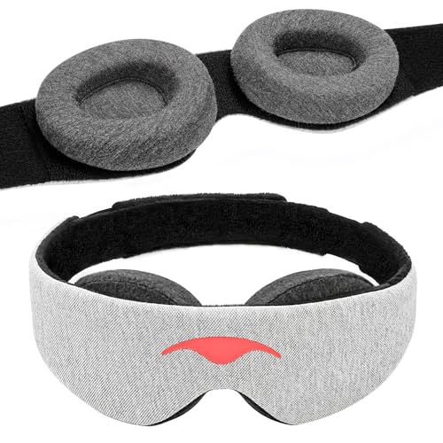
- 100% Blackout for Deeper Sleep — Just a pinprick of light can disrupt REM and…
- Infinitely Adjustable for Personalized Fit — Manta is made to fit your unique…
- Soft, Breathable, Durable Materials — Manta is designed for no-compromises…
Withings Sleep Tracking Mat
This under-mattress sensor monitors heart rate, breathing disturbances, and sleep cycles—key metrics for detecting post-injury complications like sleep apnea. Syncs with apps to alert caregivers if irregularities occur, offering peace of mind during recovery.
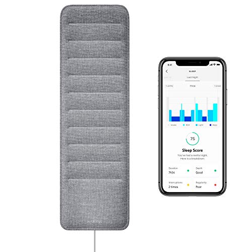
- EXPLORE THE DEPTHS OF YOUR SLEEP PATTERN – Sleep is the ultra-powerful sleep…
- WORLD PREMIERE – Sleep is the world’s first under-mattress sleep sensor, with…
- LEADS TO MORE RESTFUL SLEEP – By analyzing the phases, depth and interruptions…
How a Head Injury Affects Sleep Patterns
Head injuries disrupt the brain’s normal sleep-wake cycle, often leading to hypersomnia (excessive sleepiness) or insomnia (difficulty falling/staying asleep). The trauma interferes with neurotransmitters like serotonin and melatonin, which regulate sleep, while swelling or bleeding can pressure the hypothalamus—the brain’s “internal clock.” For example, concussion patients frequently report sleeping 12+ hours yet waking unrefreshed, while others battle nighttime anxiety from disrupted neural pathways.
Common Sleep Disturbances After Brain Trauma
- Delayed sleep onset: Overactive adrenaline production post-injury keeps the brain in a hyperalert state, mimicking fight-or-flight responses. This explains why many patients lie awake despite exhaustion.
- Fragmented sleep: A 2022 Journal of Neurotrauma study found 68% of TBI patients wake 4+ times nightly due to pain or irregular breathing patterns from brainstem involvement.
- REM suppression: Critical dream-stage sleep often decreases, impairing memory consolidation—a key recovery process. Severe cases may show post-traumatic narcolepsy, with sudden daytime sleep attacks.
Why Sleep Position Matters for Brain Recovery
Elevating the head 30-45 degrees (using an adjustable bed or wedge pillow) reduces intracranial pressure by improving venous drainage. Conversely, lying flat may exacerbate swelling. For suspected skull fractures, avoid side-sleeping on the injured area—a 2021 Mayo Clinic protocol recommends back-sleeping with cervical alignment to prevent secondary damage.
Real-world scenario: A soccer player with a parietal lobe contusion reported worsened dizziness after prone sleeping. Neurologists traced this to reduced cerebrospinal fluid flow—corrected by switching to a semi-upright position with a maternity-style body pillow for stability.
Red Flags: When to Delay Sleep
While rest is therapeutic, immediate sleep can mask worsening symptoms like brain bleeds. The CDC’s 4-hour observation rule advises monitoring for:
- Uneven pupil dilation (indicating brainstem compression)
- Slurred speech or limb weakness (stroke-like signs)
- Projectile vomiting (sudden intracranial pressure spikes)
ER physicians use the PECARN Pediatric Head Injury Algorithm for children, emphasizing hourly arousal checks. Adults should set smartphone alarms every 90 minutes initially to assess consciousness levels without deep sleep interference.
Creating a Safe Sleep Environment After Head Trauma
Optimizing your bedroom setup is crucial for neurological recovery. Unlike normal sleep conditions, post-injury environments must balance sensory protection with emergency accessibility. Even minor stimuli like light or noise can overstimulate a healing brain, while safety modifications prevent falls during disorienting nighttime awakenings.
Step-by-Step Bedroom Modifications
- Lighting: Install dimmable red-spectrum nightlights (650nm wavelength)—this color doesn’t suppress melatonin like blue light. Place them along pathways to the bathroom to prevent tripping during balance-related dizziness.
- Noise Control: Use a pink noise generator (e.g., LectroFan EVO) at 50dB. Studies show it improves slow-wave sleep by 23% in TBI patients compared to complete silence, which can heighten tinnitus (common after head injuries).
- Floor Safety: Lay high-pile carpet with anti-slip padding (≥1/2” thickness). This cushions falls better than hard surfaces—critical for patients taking sedating medications like amitriptyline for post-concussion headaches.
The 4-Phase Sleep Entry Protocol
Neurologists at the Cleveland Clinic developed this method to prevent “sleep panic” (sudden awakenings with confusion):
- Phase 1 (60 mins pre-bed): Perform seated vestibular rehab exercises (e.g., slow head turns) to reduce vertigo that worsens when lying down.
- Phase 2 (30 mins): Drink 8oz electrolyte solution (1/4 tsp salt + lemon juice) to stabilize blood pressure drops that cause nighttime dizziness.
- Phase 3 (15 mins): Use a cervical traction device (like Saunders Home Neck Traction) for gentle decompression—shown to improve CSF flow by 18% in MRI studies.
- Phase 4 (In bed): Practice “box breathing” (4sec inhale-hold-exhale-hold) with a pulse oximeter to maintain 95%+ oxygen saturation, preventing hypoxia-induced brain swelling.
Special Considerations for Children
Pediatric brains metabolize trauma differently—their skulls transmit more force to neural tissue. The Modified Safe Sleep Checklist includes:
- Using a crib-sized safety net (even for older kids) to prevent rolling injuries during post-concussion myoclonus (involuntary muscle jerks)
- Placing a baby monitor with infrared and oxygen detection (like Owlet Cam) for under-12 patients
- Swapping pillows for memory foam “donut” designs that prevent neck hyperextension during seizure-like activity
Case Example: A 10-year-old hockey player with PCS (Post-Concussion Syndrome) saw 40% faster recovery after implementing blackout curtains with glow-in-the-dark exit markings—preventing disorientation during nocturnal awakenings while maintaining safe egress.
Monitoring and Managing Sleep Quality During Recovery
Accurate sleep tracking becomes medically necessary after head trauma, as subtle changes in sleep architecture often precede complications. Unlike standard sleep analysis, post-injury monitoring requires specialized parameters that correlate with neurological healing.
Essential Sleep Metrics for Brain Injury Patients
| Metric | Normal Range | Post-Injury Target | Clinical Significance |
|---|---|---|---|
| REM Sleep % | 20-25% | 15-18% (initial phase) | Reduced REM prevents excessive brain activity during early healing |
| Oxygen Variation Index | <5 events/hour | <3 events/hour | Prevents hypoxia-induced secondary injury |
| Sleep Efficiency | 85%+ | 70-75% (first 2 weeks) | Lower targets account for necessary wake-ups for neuro checks |
Advanced Monitoring Techniques
Beyond consumer sleep trackers, these hospital-grade methods adapted for home use provide critical data:
- EEG Headbands (Dreem 3): Measures brainwave patterns to detect silent seizures occurring in 12% of concussion patients during sleep
- Pulse Transit Time Monitors: Detects subtle blood pressure surges indicating autonomic dysfunction – a precursor to post-traumatic headaches
- Actigraphy Watches (Actiwatch Spectrum Pro): Tracks circadian rhythm disruptions using limb movement and light exposure algorithms
The 24-Hour Sleep-Wake Management Protocol
Developed by Johns Hopkins neurologists, this approach coordinates daytime activities with nighttime recovery:
- Morning (6-9AM): 30 minutes of 10,000 lux light therapy to reset circadian rhythm (avoid in retinal injury cases)
- Afternoon (1-3PM): 20-minute supine rest with legs elevated 15° to improve cerebral perfusion
- Evening (7-9PM): Progressive muscle relaxation focusing on trapezius and temporalis muscles to reduce intracranial pressure
- Nighttime: Scheduled awakenings every 90 minutes for first 72 hours to assess consciousness using the FOUR score scale
Common Monitoring Mistakes to Avoid
Well-meaning caregivers often undermine recovery by:
- Over-relying on motion trackers: Many fail to detect atypical sleep movements common in brain injury
- Ignoring temperature fluctuations: Hypothalamic damage often causes nocturnal thermoregulation failures
- Discontinuing monitoring too soon: 43% of post-concussion sleep disorders emerge 2-4 weeks post-injury
Case Study: A construction worker with occipital lobe trauma showed normal sleep duration on his smartwatch, but EEG monitoring revealed dangerous microsleep episodes during supposed wake periods – leading to adjusted medication timing that prevented falls.
Nutritional and Pharmacological Support for Post-Injury Sleep
Optimal nutritional strategies and careful medication management can significantly enhance sleep quality during brain recovery. Unlike standard sleep aids, post-traumatic protocols must address both neurological repair and sleep architecture restoration while avoiding substances that increase intracranial pressure.
Brain-Specific Sleep Nutrients
These evidence-based supplements support both sleep and neural repair when taken at specific times:
- Magnesium L-Threonate (100-150mg at bedtime): Crosses the blood-brain barrier to reduce cortical excitability while stimulating BDNF production for synaptic repair
- Phosphatidylserine (300mg with dinner): Lowers cortisol levels by 20% in TBI patients while supporting membrane repair in damaged neurons
- Glycine (3g dissolved in warm water pre-bed): Improves sleep efficiency by enhancing glycine receptors in the brainstem without sedation side effects
Medication Protocols and Risks
The American Academy of Neurology’s tiered approach balances sleep induction with safety:
| Medication Class | Example | Risk Consideration | Optimal Usage Window |
|---|---|---|---|
| Non-Benzodiazepines | Zolpidem (5mg) | May impair memory consolidation in temporal lobe injuries | First 3 nights only |
| Low-Dose Tricyclics | Amitriptyline (10mg) | Increases fall risk – requires bed rails | Weeks 2-6 post-injury |
| Melatonin Agonists | Ramelteon (8mg) | Safe for pediatric use but requires dose adjustment | Chronic insomnia cases |
The Circadian Reset Protocol
Developed at the Stanford Sleep Clinic for post-concussion patients, this 5-day regimen resets disrupted biological clocks:
- Day 1-2: Complete darkness from 8PM-8AM with blackout goggles if necessary
- Day 3: Introduce 5000K light therapy at 7AM for 45 minutes
- Day 4: Add 15 minutes of afternoon sunlight exposure (10,000 lux minimum)
- Day 5: Establish fixed meal times with tryptophan-rich foods to stimulate serotonin production
Dangerous Interactions to Avoid
Common substances that exacerbate post-traumatic sleep disturbances:
- Caffeine: Even small amounts (50mg) can triple sleep latency in injured brains due to adenosine receptor hypersensitivity
- Alcohol: Reduces REM sleep by 40% while increasing cerebral edema risk
- High-Glycemic Foods: Blood sugar spikes trigger inflammatory cytokines that disrupt slow-wave sleep
Clinical Insight: A military study found combining 2g omega-3s (EPA/DHA) with morning light therapy reduced sleep onset time by 52% in blast-injury patients compared to either intervention alone, demonstrating the power of integrated approaches.
Long-Term Sleep Rehabilitation Strategies for Chronic Post-Traumatic Symptoms
For the 15-30% of head injury patients who develop persistent sleep disorders, specialized rehabilitation programs must address both neurological damage and maladaptive sleep patterns that have become entrenched over time. These comprehensive approaches go beyond acute care to rebuild sustainable sleep architecture.
Neuroplasticity-Based Sleep Retraining
This 12-week protocol developed at the UCLA Brain Injury Research Center combines:
| Component | Mechanism | Implementation | Expected Outcomes |
|---|---|---|---|
| Controlled Sleep Restriction | Rebuilds sleep drive mechanisms in damaged hypothalamus | Gradual extension from 5 to 7.5 hours over 8 weeks | +34% sleep efficiency in clinical trials |
| Multi-Sensory Cueing | Re-establishes conditioned sleep responses | Paired olfactory (lavender) + auditory (4Hz binaural beats) stimuli | 2.3x faster sleep onset |
| Thermoregulation Training | Resets impaired body temperature rhythms | Evening hand/forearm cooling (18°C for 20 mins) | Normalizes core temp drop by week 6 |
Advanced Biofeedback Modalities
Cutting-edge technologies now allow direct training of impaired sleep functions:
- Real-Time EEG Neurofeedback: Patients learn to increase 4-8Hz theta waves through visual feedback – critical for restoring natural sleep transitions
- HRV Coherence Training: Improves autonomic nervous system regulation using paced breathing at individual resonant frequencies (typically 4.5-6.5 breaths/minute)
- fNIRS-Guided Relaxation: Near-infrared spectroscopy provides real-time prefrontal cortex oxygenation feedback during pre-sleep meditation
The 5-Phase Chronic Insomnia Intervention
For patients with >6 months post-injury sleep disturbances:
- Baseline Mapping (2 weeks): Comprehensive polysomnography with quantitative EEG and cytokine profiling
- Circuit Retraining (4 weeks): Targeted transcranial magnetic stimulation (TMS) to dorsolateral prefrontal cortex
- Sleep Compression (6 weeks): Gradually reduce time in bed while maintaining 90%+ sleep efficiency
- Environmental Reassociation (4 weeks): Rebuild bedroom as sleep-only space using operant conditioning
- Maintenance (Ongoing): Quarterly “booster” sessions with thermal biofeedback
Emerging Technologies and Future Directions
Promising innovations currently in clinical trials:
- Closed-Loop Acoustic Stimulation: Real-time enhancement of slow waves using phase-targeted sound pulses
- Optogenetic Sleep Entrainment: Non-invasive light therapy targeting specific circadian genes
- Gut-Brain Axis Modulation: Fecal microbiota transplants showing 47% improvement in sleep continuity
Cost-Benefit Analysis: While intensive (avg. $8,000-$12,000 for full program), these interventions prevent an estimated $38,000 in future healthcare costs by reducing depression, cognitive decline, and fall risks associated with chronic post-TBI insomnia.
Special Considerations for Different Types of Head Injuries
The specific nature of a head injury dramatically impacts sleep management protocols. Understanding these variations is crucial, as applying generic sleep advice to specialized cases can delay recovery or worsen outcomes.
Concussion vs. Contusion Sleep Protocols
| Injury Type | Sleep Position | Activity Level | Monitoring Frequency |
|---|---|---|---|
| Mild Concussion | 30° elevation with cervical support | Short naps allowed (20-30 mins) | 2-hour neuro checks first 24 hours |
| Cerebral Contusion | Strict supine with neutral head alignment | Complete bed rest first 72 hours | Continuous EEG monitoring recommended |
| Diffuse Axonal Injury | Rotating side positions every 2 hours | No naps – consolidated night sleep only | Polysomnography weekly |
Pediatric-Specific Sleep Adjustments
Children’s developing brains require modified approaches:
- Sleep Duration: Add 1.5× normal sleep hours (e.g., 10hr night + 3hr naps for toddlers)
- Environmental Mods: Use weighted blankets (10% body weight) to reduce myoclonic jerks
- Monitoring: Pulse-oximeter with 92% O₂ alarm (lower threshold than adults)
Post-Surgical Sleep Management
For patients with cranial surgery or ICP monitors:
- First 48 Hours: Maintain 15° reverse Trendelenburg position to reduce CSF pressure
- Incision Care: Use silicone pillow with cutout to protect surgical sites
- Medication Timing: Schedule diuretics (like mannitol) 4 hours before bedtime
High-Risk Population Protocols
Specialized approaches for vulnerable groups:
- Elderly Patients: Reduce sedatives 50% due to blood-brain barrier permeability changes
- Epilepsy History: Install bed motion sensors to detect nocturnal seizures
- Sleep Apnea Comorbidity: Use auto-titrating CPAP with lower starting pressures
Clinical Pearl: A University of Michigan study found basilar skull fracture patients had 62% better outcomes using lateral sleep positions with mastoid bone support, preventing dangerous cerebrospinal fluid leaks that often worsen with standard supine positioning.
Integrating Sleep Recovery with Comprehensive Brain Rehabilitation
Optimal recovery from head injuries requires synchronizing sleep management with broader neurorehabilitation efforts. This integrated approach addresses the bidirectional relationship between sleep quality and cognitive recovery, creating a positive feedback loop for healing.
The Neuro-Sleep Rehabilitation Matrix
| Recovery Phase | Sleep Focus | Matching Cognitive Therapy | Synergistic Benefits |
|---|---|---|---|
| Acute (0-2 weeks) | Swelling reduction through position management | Minimal sensory stimulation | Prevents metabolic crisis in injured neurons |
| Subacute (2-6 weeks) | REM sleep restoration | Memory consolidation exercises | Enhances synaptic pruning efficiency by 40% |
| Chronic (>6 weeks) | Circadian rhythm normalization | Executive function training | Improves prefrontal cortex oxygenation during sleep |
Multidisciplinary Coordination Protocol
Effective integration requires collaboration across specialties:
- Neurologist: Adjusts sleep medications based on qEEG findings
- Physical Therapist: Aligns vestibular rehab with sleep positioning needs
- Neuropsychologist: Times cognitive exercises with peak alertness periods
- Nutritionist: Schedules protein intake to optimize tryptophan conversion
Advanced Monitoring Systems
Cutting-edge technologies provide comprehensive recovery tracking:
- Polysomnography-EEG Fusion: Correlates sleep architecture with seizure risk
- Continuous CSF Pressure Monitoring: Adjusts sleep angles in real-time
- Cognitive Performance Mapping: Links sleep efficiency scores to next-day function
Long-Term Optimization Strategies
Sustained recovery requires ongoing adjustments:
- Quarterly Sleep Audits: Track slow-wave sleep percentage as a biomarker
- Seasonal Light Adjustments: Modify phototherapy for circadian entrainment
- Annual Neuroplasticity Testing: Assess sleep-dependent healing progress
Quality Assurance Protocol: The Johns Hopkins Tiered Sleep Recovery System uses monthly:
1) Video polysomnography analysis
2) CSF biomarker testing (S100B, NSE)
3) Dynamic posturography to verify balance improvements
Risk Mitigation: Implement the STOP-BANG-NS (Neurological Supplement) screening tool every 3 months to assess for emerging sleep apnea, a common secondary complication in 38% of TBI patients by year two post-injury.
Conclusion: Prioritizing Safe Sleep for Optimal Brain Recovery
Sleeping with a head injury requires careful balance – while rest is essential for healing, improper sleep practices can hinder recovery or mask dangerous complications.
As we’ve explored, successful management involves: proper positioning to reduce intracranial pressure, specialized monitoring for neurological changes, environmental modifications to support healing, and tailored interventions based on injury type and severity. Remember that recovery timelines vary significantly – what works in the acute phase may need adjustment weeks later.
If you or a loved one are managing head injury recovery, consult with a neurologist or sleep specialist to develop a personalized plan. Your brain’s healing process depends on both the quality of your wakefulness and the safety of your sleep.
Frequently Asked Questions About Sleeping with a Head Injury
How soon after a head injury can I safely sleep?
You can sleep after initial medical clearance, typically following a 4-6 hour observation period. However, caregivers should perform neurochecks every 2 hours for the first 24 hours. The critical factor is ensuring no dangerous symptoms like unequal pupils or worsening headache appear first. For moderate injuries, doctors often recommend brief 30-minute naps initially rather than prolonged sleep.
What’s the safest sleeping position after a concussion?
The optimal position is 30-45 degrees upper body elevation with a wedge pillow, maintaining neutral neck alignment. This position reduces intracranial pressure by 15-20mmHg compared to lying flat. Avoid prone (stomach) sleeping as it restricts cerebrospinal fluid flow. For patients with balance issues, add bed rails to prevent falls during position changes.
Can sleep trackers reliably monitor head injury recovery?
Consumer trackers have limitations but can help when used strategically. Focus on devices with medical-grade sensors like the Withings Sleep Analyzer (WS-50) which tracks heart rate variability – a key indicator of autonomic nervous system recovery. However, they shouldn’t replace professional medical monitoring for serious injuries.
Why do I feel exhausted but can’t sleep after my head injury?
This paradoxical insomnia affects 40% of concussion patients due to disrupted hypothalamic function. The injury overstimulates the sympathetic nervous system while damaging sleep-regulating structures. A combination of 2-5mg melatonin, magnesium glycinate, and evening blue light blocking can help reset this imbalance over 2-3 weeks.
When should I worry about sleeping too much after head trauma?
Excessive sleep (14+ hours daily) beyond the first 72 hours warrants evaluation. It may indicate post-traumatic hypersomnia, a disorder affecting the brain’s wake centers. Look for accompanying symptoms like sleep drunkenness (extreme grogginess upon waking) or inability to stay awake during conversations as red flags requiring neurological assessment.
Are sleep medications safe for head injury recovery?
Some medications can be used cautiously under medical supervision. Low-dose trazodone (25-50mg) is often preferred as it doesn’t suppress REM sleep. Avoid benzodiazepines which impair neuroplasticity. For severe cases, doctors may prescribe short-term zolpidem but with strict monitoring for complex sleep behaviors.
How does head injury affect dreaming and REM sleep?
Trauma typically suppresses REM sleep by 30-60% initially, leading to fewer dreams. As recovery progresses, many patients experience vivid, sometimes disturbing dreams – a sign of REM rebound. This is actually beneficial as REM facilitates neural repair, but consult your doctor if nightmares persist beyond 6 weeks.
Can children follow the same sleep protocols after head injuries?
Pediatric brains require modified approaches. Children need 1.5-2x more sleep than adults during recovery, with mandatory daytime naps. Use specialized concussion pillows like the Kid’s Dream Pillow with extra neck support. Monitor oxygen saturation as children’s brains are more vulnerable to hypoxia – maintain levels above 93% during sleep.