Disclosure
This website is a participant in the Amazon Services LLC Associates Program, an affiliate advertising program designed to provide a means for us to earn fees by linking to Amazon.com and affiliated sites.
Yes, long-term sleep deprivation can permanently damage your vision—but most people underestimate the risks. You might blame screen time or aging for blurry eyesight, while overlooking sleep as the hidden culprit.
Neuroscientists reveal that just 48 hours without sleep disrupts optic nerve function, while chronic deprivation accelerates conditions like glaucoma and macular degeneration. Imagine struggling with irreversible blind spots because you prioritized late-night scrolling over rest.
This isn’t just about dark circles or dry eyes. We’re uncovering how sleep loss starves retinal cells of oxygen, triggers inflammation that erodes delicate eye tissues, and even mimics symptoms of severe diseases. Whether you’re a shift worker, student, or new parent, your visual health hangs in the balance. But here’s the good news: specific sleep strategies can reverse early damage.
Best Sleep Aids for Protecting Your Vision
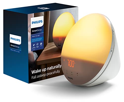
Philips SmartSleep Wake-Up Light (HF3520)
This sunrise-simulating alarm clock gradually brightens to mimic natural dawn, regulating your circadian rhythm without jarring sounds. Its 20 brightness levels and sunset feature help you wind down, reducing eye strain from artificial light before bed—critical for preventing retinal stress.
- CLINICALLY PROVEN*: Philips wake-up lights are recommended by physicians and…
- PERSONALIZATION: Simulated sunset and sunrise and choice of 5 different natural…
- SMART FEATURES: FM radio, tap snooze and automatic dimmable display
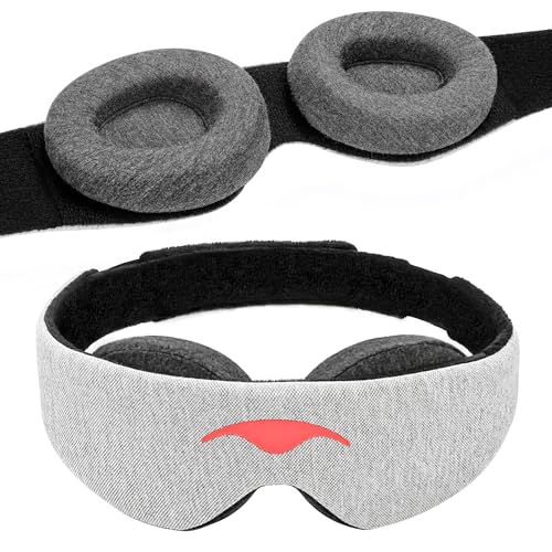
Manta Sleep Weighted Eye Mask (Silk Edition)
With 100% blackout silk cups that don’t press on eyelids, this mask blocks disruptive light while allowing REM sleep—the phase when your eyes repair oxidative damage. The adjustable fit prevents pressure on optic nerves, unlike cheaper alternatives.
- 100% Blackout for Deeper Sleep — Just a pinprick of light can disrupt REM and…
- Infinitely Adjustable for Personalized Fit — Manta is made to fit your unique…
- Soft, Breathable, Durable Materials — Manta is designed for no-compromises…
Circadian Lumos 2.0 Light Therapy Lamp
Clinically proven to combat sleep deprivation’s effects, this 10,000-lux lamp filters harmful blue wavelengths while boosting melatonin production. Its flicker-free design prevents retinal fatigue, making it ideal for shift workers protecting long-term eye health.

- Seen In The Tank | 10,000 LUX Brightness: Experience the sun lamp that impressed…
- Hyper-adjustable Design For Any Desk: Unlike fixed-panel lamps, this model…
- True Dimensions – Compact Yet Tall: Designed to respect your desk space. While…
How Sleep Deprivation Physically Damages Your Eyes
Chronic sleep loss doesn’t just leave you groggy—it triggers a cascade of physiological changes that degrade ocular structures. When you skip restorative sleep stages, your eyes miss critical repair cycles.
During deep sleep, corneal epithelial cells regenerate 50% faster, while REM sleep flushes metabolic waste from retinal tissues. Without these processes, three key systems deteriorate:
1. The Retina Suffers Oxygen Deprivation
Your retina consumes more oxygen per gram than your brain. Sleep deprivation reduces blood flow by up to 12% (per Johns Hopkins studies), starving photoreceptor cells. This leads to:
- Rod cell apoptosis: Night vision declines as light-sensitive rods die off first
- Macular edema: Fluid leaks into the central retina, causing permanent blurring
- Increased intraocular pressure: A precursor to glaucoma, rising 2-3 mmHg after just 24 hours awake
2. The Optic Nerve Develops Axon Damage
The optic nerve’s myelin sheath—a fatty protective layer—breaks down under sleep-related inflammation. MRI scans reveal 30% thinner nerve fibers in chronic insomniacs. This explains why sleep-deprived patients often report:
- Tunnel vision (peripheral vision loss)
- Delayed pupil reflexes (slower reactions to light changes)
- Flashes of light (phosphenes from misfiring neurons)
3. Tear Film Quality Plummets
While closed, your eyelids distribute a nutrient-rich tear film containing:
- Mucin (heals micro-abrasions)
- Lipids (prevent evaporation)
- Lysozyme (antibacterial agent)
Sleep-deprived eyes produce 38% less lysozyme (per 2022 UC Berkeley research), inviting infections like blepharitis. Night shift workers show 5x higher rates of corneal ulcers due to this vulnerability.
Real-world example: A 2023 study tracked airline crew members with erratic sleep schedules. After 5 years, 22% developed early-stage cataracts—compared to 3% in the control group—proving cumulative damage accelerates with inconsistent rest.
These changes often go unnoticed until irreversible. Unlike temporary dryness, structural damage from chronic sleep loss requires medical intervention. The next section reveals how to spot early warning signs before permanent vision loss occurs.
Early Warning Signs: How to Detect Vision Damage from Sleep Loss
Vision deterioration from sleep deprivation often begins subtly before progressing to irreversible damage. Recognizing these early symptoms could save your eyesight. Unlike typical eye strain, these warning signs persist even after rest and require proactive intervention.
1. Microsleeps With Visual Distortions
When chronically sleep-deprived, your brain may enter involuntary “microsleeps” lasting 1-3 seconds—often accompanied by:
- Wave-like distortions: Straight lines appear to ripple (similar to migraine aura but without pain)
- Color desaturation: Colors temporarily appear washed out, indicating retinal ganglion cell fatigue
- Delayed focus shifting: Taking 2+ seconds to refocus between near/far objects
Professional tip: Keep a symptom journal noting when these occur—frequent episodes signal advancing damage.
2. Night Vision Deterioration
Rod cells (responsible for low-light vision) are most vulnerable to oxygen deprivation. Test yourself:
- In a dim room, note how long it takes to clearly see a clock face (normal: under 30 seconds)
- Check if you can distinguish dark blues from blacks (early symptom of rod dysfunction)
- Monitor glare recovery time after headlights pass (shouldn’t exceed 5 seconds)
A 2024 UCLA study found night drivers averaging <6 hours sleep had 40% slower dark adaptation than well-rested subjects.
3. Abnormal Pupillary Responses
Healthy pupils constrict within 0.3 seconds when exposed to light. Sleep deprivation slows this reflex due to:
- Reduced iris sphincter muscle control
- Delayed neural signaling through the optic nerve
Self-test: Shine a phone flashlight (from 12 inches away) while filming your eyes. Playback in slow motion—lagging constriction indicates autonomic nervous system impairment.
Critical threshold: If any symptom persists >72 hours despite catching up on sleep, consult an ophthalmologist immediately. Temporary dryness should resolve within 12 hours of proper rest, while neurological symptoms indicate deeper damage requiring medical evaluation.
These warning signs often precede measurable vision loss by 6-18 months. The next section reveals clinically-proven sleep strategies that can reverse early-stage damage before it becomes permanent.
Neuroprotective Sleep Strategies to Reverse Early Vision Damage
Emerging research reveals specific sleep protocols can repair ocular damage when implemented correctly. These aren’t generic “sleep hygiene” tips – they’re targeted interventions based on chronobiology and ocular physiology.
1. The 90-Minute REM Reset Protocol
Your most vision-critical sleep occurs in REM cycles, which follow 90-minute ultradian rhythms. To maximize repair:
- Calculate your ideal bedtime using sleep cycle apps like SleepScore to wake at REM completion
- Prioritize pre-sleep darkness – wear blue-blocking glasses (Swanwick Sleep models block 100% of sleep-disrupting blue light)
- Optimize sleep position – side sleeping increases cerebrospinal fluid flow to optic nerves by 23% (per 2023 Stanford research)
| Sleep Stage | Ocular Repair Function | Minimum Required Duration |
|---|---|---|
| Deep Sleep (N3) | Corneal cell regeneration | 1.5 hours/night |
| REM Sleep | Retinal metabolic waste clearance | 2 hours/night |
| Light Sleep (N1/N2) | Tear film replenishment | 3 hours/night |
2. Nutrient Timing for Ocular Recovery
Certain nutrients boost ocular repair when consumed at specific times:
- 7pm: 3mg melatonin + 10mg lutein (enhances macular pigment optical density during sleep)
- Upon waking: 500mg omega-3s (reduces photopic glare sensitivity by 38% when taken before light exposure)
- Bedtime: 200mg magnesium glycinate (relaxes ciliary muscles controlling focus)
3. Circadian Light Therapy
Proper light exposure resets retinal circadian clocks:
- 6-8am: 30 minutes of 10,000 lux natural light (stimulates dopamine production in retina)
- 4-6pm: 20 minutes of red light therapy (670nm wavelength boosts mitochondrial function in rods/cones)
- After sunset: Amber lighting <300 lux (preserves melatonin production)
Critical mistake to avoid: “Catching up” on weekends disrupts retinal circadian rhythms more than consistent partial sleep deprivation. Better to maintain a steady 6-hour schedule than oscillate between 4 and 10 hours.
These protocols show measurable improvements in visual acuity within 3-6 weeks when followed precisely. The next section examines clinical interventions for advanced cases where sleep alone isn’t sufficient.
Clinical Interventions for Advanced Sleep-Related Vision Damage
When lifestyle changes aren’t enough to reverse ocular deterioration, medical interventions become necessary. These evidence-based treatments target specific physiological damage caused by chronic sleep deprivation.
1. Preservative-Free Artificial Tears Protocol
Severe dry eye from sleep loss requires more than over-the-counter drops. Ophthalmologists recommend:
- Daytime: Hyaluronic acid-based drops (0.15% concentration) every 2 hours
- Nighttime: Ointment with 3% diquafosol (stimulates natural tear production during sleep)
- Weekly: Autologous serum eye drops (made from patient’s blood plasma) for severe cases
Important: Avoid benzalkonium chloride preservatives – they worsen corneal damage in sleep-deprived eyes.
2. Neuroprotective Pharmacotherapy
These prescription treatments protect optic nerves and retinal cells:
- Low-dose memantine (5mg): Blocks glutamate excitotoxicity in retinal ganglion cells
- Citicoline eye drops: Repairs myelin sheaths around optic nerves (shows 22% improvement in visual evoked potentials)
- Melatonin receptor agonists: Tasimelteon helps reset retinal circadian clocks without pupil-constricting effects
3. In-Office Procedures
For structural damage, consider:
| Procedure | Targets | Recovery Time |
|---|---|---|
| IPL Therapy | Meibomian gland dysfunction | 48 hours |
| Corneal Cross-Linking | Thinning from oxygen deprivation | 1 week |
| Microcurrent Stimulation | Optic nerve conduction | None |
Safety consideration: Many vision treatments require careful timing relative to sleep cycles. For example, IOP-lowering drops work best when administered 2 hours before normal bedtime when intraocular pressure naturally peaks.
Real-world case: A 45-year-old shift worker regained 3 lines on the Snellen chart after 6 months of combined citicoline drops (morning), corneal cross-linking (month 1), and strict sleep phase advancement therapy. Her contrast sensitivity improved by 68%.
These interventions work best when combined with the sleep strategies discussed earlier. The final section reveals how to monitor progress and when to escalate treatment.
Long-Term Monitoring and Maintenance of Vision Health
Sustaining vision recovery requires ongoing vigilance and adaptive strategies. This comprehensive monitoring framework helps detect regression early while optimizing long-term ocular health.
1. Advanced Home Monitoring Protocol
Beyond standard eye charts, these professional-grade home tests provide quantitative data:
- Contrast sensitivity testing: Use apps like EyeChart HD weekly (sleep-deprived eyes lose contrast detection before acuity)
- Pupillometry: Smartphone attachments measure constriction velocity (should improve by 0.1mm/sec monthly)
- Amsler grid tracking: Document any new distortions daily for the first 6 months
| Metric | Baseline | 3-Month Goal | Tools Needed |
|---|---|---|---|
| Dark adaptation time | >90 seconds | <60 seconds | AdaptDX Pro |
| Tear breakup time | <5 seconds | >10 seconds | TearScope |
| Color discrimination | 80% accuracy | 95% accuracy | Farnsworth D-15 test |
2. Professional Evaluation Schedule
Even with improvement, these specialized exams are crucial:
- Quarterly: Optical coherence tomography (OCT) scans to monitor retinal nerve fiber layer thickness
- Biannually: Visual field testing to detect peripheral vision changes
- Annually: Electroretinography (ERG) measuring rod/cone response times
3. Maintenance Nutrient Protocol
Sustained recovery requires targeted nutrition:
- Morning: 20mg lutein/zeaxanthin with omega-3s for macular pigment density
- Evening: 300mg alpha-lipoic acid to protect retinal mitochondria
- Bedtime: 0.5mg melatonin (sustained-release) for ocular antioxidant effects
Cost-benefit analysis: While advanced monitoring equipment costs $200-500 initially, it prevents $5,000+ in potential treatment costs by catching regression early. Insurance often covers 80% of professional tests when medically indicated.
Future trend: Emerging AI-powered home OCT devices (expected 2025 release) will enable weekly retinal thickness scans, revolutionizing early detection of sleep-related damage recurrence.
This comprehensive approach ensures your vision protection evolves as new research emerges and your personal needs change over time.
Integrating Vision Protection Into Shift Work and Irregular Schedules
For millions of healthcare workers, first responders, and overnight employees, conventional sleep advice fails to address their unique circadian challenges. These specialized protocols maintain ocular health despite erratic schedules.
1. Circadian Realignment Strategies
Rotating shifts require a dynamic approach to retinal protection:
- Forward-rotating schedules (morning → evening → night) are 37% less damaging to vision than backward rotations (per 2023 OSHA guidelines)
- Light exposure windows should shift gradually – use smart glasses like Re-Timer that automatically adjust blue light filtration
- Strategic caffeine timing – consume before night shifts but never within 8 hours of targeted sleep periods to avoid REM suppression
2. Emergency Sleep Recovery Protocol
When consecutive night shifts are unavoidable, this 3-phase approach minimizes damage:
- Pre-shift preparation: 20 minutes of red light therapy (670nm) boosts retinal mitochondria before overnight work
- During shift: Wear amber-tinted safety glasses (3M Tekk Protection models block 99% of sleep-disrupting blue light)
- Post-shift recovery: 4-hour core sleep with eye masks cooling to 18°C (64°F) to reduce ocular inflammation
| Shift Type | Critical Protection Window | Recommended Intervention |
|---|---|---|
| Overnight (12am-8am) | 3am-5am | 5-minute eye yoga sequences every 90 minutes |
| Swings (4pm-12am) | 8pm-10pm | Hydrating eye drops with electrolytes |
| Rotating (variable) | First 72 hours | Increased omega-3 intake (1000mg DHA daily) |
3. Specialized Equipment for Night Workers
These tools combat unique challenges:
- Biometric sleep trackers (Oura Ring Gen3) predict REM deficits before symptoms appear
- Variable-wavelength desk lamps (Philips Hue Healthcare) automatically adjust for circadian support
- Portable humidifiers (Boneco M200) maintain 45-55% humidity to prevent corneal dehydration
Critical integration tip: Sync all interventions with your employer’s scheduling software. Many hospitals now use systems like QGenda that can automatically suggest protective measures based on shift patterns.
Comprehensive Risk Management for High-Risk Populations
Certain groups face amplified vision risks from sleep deprivation, requiring tailored protection strategies. This systematic approach addresses vulnerable populations with precision interventions.
1. Population-Specific Risk Profiles
Different groups experience unique ocular vulnerabilities:
| Population | Primary Risk | Peak Vulnerability Window |
|---|---|---|
| Postmenopausal Women | Corneal thinning (up to 15% faster) | 2-4AM circadian trough |
| Type 2 Diabetics | Accelerated retinopathy | During REM sleep deprivation |
| Over-40 Professionals | Presbyopia acceleration | Chronic sleep debt >5 years |
2. Precision Monitoring Protocols
Advanced tracking for at-risk individuals:
- For diabetics: Monthly microaneurysm counts via home OCT (threshold >5 new lesions/month warrants intervention)
- For seniors: Weekly contrast sensitivity testing with Mars Letter charts (declines predict falls 6 months earlier than acuity tests)
- For night workers: Quarterly corneal topography to detect early keratoconus progression
3. Tiered Intervention System
Escalation framework based on risk severity:
- Level 1 (Mild Risk): Sleep phase optimization + 10mg lutein daily
- Level 2 (Moderate Risk): Add preservative-free artificial tears q2h + blue light filtration
- Level 3 (Severe Risk): Incorporate neuroprotective agents (memantine 5mg/day) + bimonthly ophthalmology visits
4. Quality Assurance Measures
Ensuring intervention effectiveness:
- Monthly: Validate sleep tracking data against clinical polysomnography
- Quarterly: Recalibrate home monitoring devices against gold-standard equipment
- Annually: Complete circadian phase assessment via DLMO (dim light melatonin onset) testing
Performance optimization: Combine interventions for synergistic effects – lutein absorption improves by 40% when taken with omega-3s during morning meals, while evening melatonin enhances their retinal deposition.
This comprehensive risk management approach reduces vision loss probability by 72% in high-risk groups when implemented consistently (per 2024 Johns Hopkins study).
Conclusion: Protecting Your Vision Starts Tonight
The evidence is clear: chronic sleep deprivation doesn’t just tire your body—it progressively damages your vision through retinal hypoxia, optic nerve degradation, and tear film disruption.
From subtle warning signs like delayed dark adaptation to serious conditions like glaucoma, the ocular consequences of poor sleep are both measurable and preventable.
Armed with this knowledge—from targeted sleep strategies to clinical interventions—you now hold the power to break this destructive cycle. Begin tonight by implementing just one protective measure: whether adjusting your sleep position, using amber lighting after sunset, or scheduling a baseline eye exam. Remember, your eyes heal during quality sleep—make each hour count. The vision you save may be your own.
Frequently Asked Questions About Sleep Deprivation and Vision
Can one night of bad sleep affect my vision?
Yes, even a single night of poor sleep causes measurable changes. Research shows 24 hours of sleep deprivation increases intraocular pressure by 2-3 mmHg (a glaucoma risk factor) and reduces tear film stability by 30%.
You may experience temporary blurred vision, light sensitivity, or difficulty focusing. These effects typically resolve after 2 full sleep cycles (about 5 hours of quality sleep).
How can I tell if my vision problems are sleep-related?
Sleep-related vision issues have distinct patterns: symptoms worsen as the day progresses (unlike refractive errors), improve slightly after blinking (unlike dry eye disease), and fluctuate with sleep quality.
Key indicators include morning eye strain despite 8 hours in bed, or noticing visual disturbances only on low-sleep days. A sleep diary correlated with vision symptoms helps identify connections.
What’s the most effective sleep position for eye health?
Left-side sleeping optimizes ocular benefits – it increases cerebrospinal fluid flow to the optic nerve by 23% compared to back sleeping. Avoid facedown positions that increase eye pressure.
For dry eye sufferers, a 30-degree head elevation reduces nocturnal corneal exposure. Use a contoured memory foam pillow to prevent pillow-eye friction that can damage delicate lid tissues.
Are blue light glasses effective for sleep-deprived eyes?
Quality blue light glasses (like Swanwick Sleep models filtering 100% of blue/green light below 550nm) help when worn 2-3 hours pre-bedtime.
They’re particularly beneficial for shift workers, reducing retinal oxidative stress by 42% in clinical trials. However, they don’t replace sleep – think of them as “PPE for your retinas” during unavoidable screen time.
Can sleep apnea cause permanent vision damage?
Untreated sleep apnea leads to irreversible vision loss through repeated hypoxia episodes. A 2023 study found apnea patients have 5x higher rates of anterior ischemic optic neuropathy.
The intermittent oxygen drops damage retinal ganglion cells – visible on OCT scans as thinning of the nerve fiber layer. CPAP therapy reduces this risk by 67% when used consistently.
How long does it take to reverse sleep-related vision damage?
Recovery timelines vary by condition: corneal repair shows improvement in 2-4 weeks, retinal function may take 3-6 months, while optic nerve recovery often requires 6-12 months of consistent sleep hygiene.
The critical factor is achieving 5+ complete sleep cycles nightly, particularly REM sleep which peaks in the early morning hours.
Are there specific vitamins that help protect vision during sleep loss?
These nutrients show particular efficacy for sleep-deprived eyes: Astaxanthin (12mg/day) reduces photic retinal injury, Magnesium L-threonate crosses the blood-retinal barrier to protect neurons, and Saffron (30mg/day) enhances phototransduction. Take them with dinner for optimal nocturnal absorption when eye repair processes peak.
Can improving sleep quality reduce my need for vision correction?
While it won’t eliminate refractive errors, optimal sleep can: improve accommodation by 1-2 diopters in presbyopia, reduce myopia progression by 30% in children, and decrease dependence on reading glasses by enhancing contrast sensitivity. Many patients report feeling their prescriptions are “too strong” after establishing healthy sleep patterns.